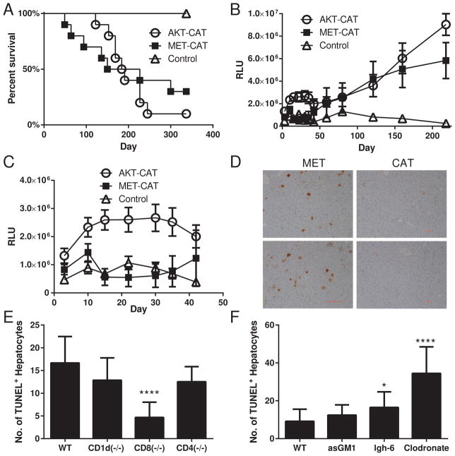
Fig. 1.
Morbidity analysis of mice with AKT+CAT and MET+CAT- initiated liver tumors using G.luc integrated in the liver. G.luc (4ug), AKT (20ug)+CAT (20ug) and MET (20ug)+CAT (20ug) oncogenes along with HSB2 (4.4ug) were delivered to C57/Bl6 mice using hydrodynamic tail vein injection. (A) Kaplan-Meier (KM) survival plot. (B) Serum luciferase levels days 0–218. (C) Serum luciferase levels days 0–42. PT3 n=5, AKT+CAT n=10, MET+CAT n=10, mean ± SEM. (D) Liver sections were ApopTag Peroxidase stained using the TUNEL method on day 7 following initiation with MET or CAT oncogenes. (E,F) MET was hydrodynamically injected into WT, CD1d−/−, CD8−/−, CD4 −/− or lgh-6 mice. For NK cell and macrophage depletion (F) mice were treated with asGM1 or Clodronate respectively on days −3 and −1 prior to MET delivery. On day 7 liver tissue sections from (E,F) were stained for TUNEL counted on 5 non-overlapping 100X fields from 3 individual mice per group using CellProfiler. n=3, ***p<0.0001 mean ± SEM. Mann-Whitney U test was used to determine significance.

