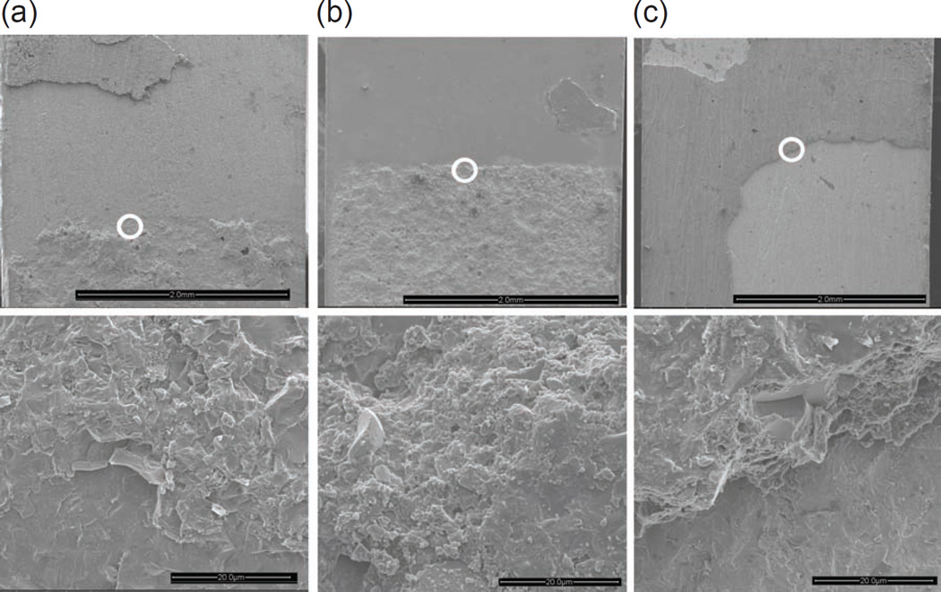
Figure 5.
SEM micrographs of the fractured surfaces of feldspathic ceramic (a), graded zirconia (b) and zirconia (c) bonded dental ceramic specimens, all subjected to sandblasting and thermocycling/aging treatments. The images conclusively correspond to a region slightly ahead of the edge of the crack starter, where slow crack growth followed by a rapid one occurred. The lower images are magnified views of the circled areas in the upper images.

