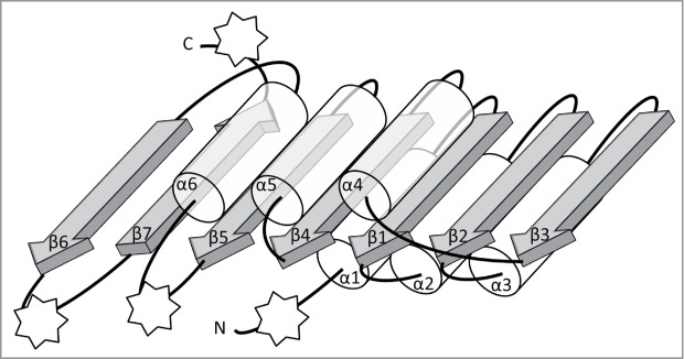
Figure 1.

Schematic representation of the conserved core of Rossmann-fold Methyltransferase (RFM) catalytic domains. The β-sheet is composed of 7 β-strands (gray arrows) surrounded by 6 α-helices (semi-transparent tubes) forms the fold that is typical for SAM-dependent methyltransferases. All secondary structure elements of the conserved core are labeled as α1, β1, etc. The stars indicate points of most frequent insertions and terminal fusions with other domains.
