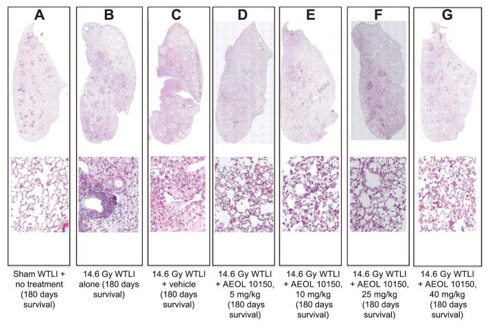
FIG. 7.
Comparison of histopathological damage among representative surviving CBA/J mice (180 days after WTLI) from different treatment groups. The top panels represent the whole-lung tissue and the bottom panels represent a magnified portion of the whole lung. Masson’s trichrome staining of lung tissue shows apparent variation in histopathology between the sham-irradiated (0 Gy) and WTLI (14.6 Gy) groups [WTLI alone, WTLI-vehicle (saline) and WTLI-AEOL 10150]. Mice treated with AEOL 10150 show less lung tissue damage compared to vehicle or nontreated mice. Panel A: Lung tissue from a sham-irradiated mouse with no treatment appears normal, lung weight (LW) 150 mg. Panel B: Lung tissue from a WTLI-alone mouse displays increased alveolar wall thickness, congestion and lymphocytic inflammation, LW 223 mg. Panel C: Lung tissue from a WTLI-vehicle mouse shows increased alveolar wall thickness, congestion and inflammation, LW 173 mg. In the lung tissue from irradiated mice treated with AEOL 10150, relatively lower alveolar wall thickness, congestion and inflammation were seen with doses of: 5 mg/kg, LW 213 mg (panel D); 10 mg/kg, LW 180 mg (panel E); 25 mg/kg, LW 206 mg (panel F); and 40 mg/kg, LW 183 mg (panel G). Since CBA/J mice are prone to pneumonitis instead of fibrosis, even WTLI cohorts display relatively scant collagen staining (blue color). Instead, the changes are suggestive of representing an organized exudate.

