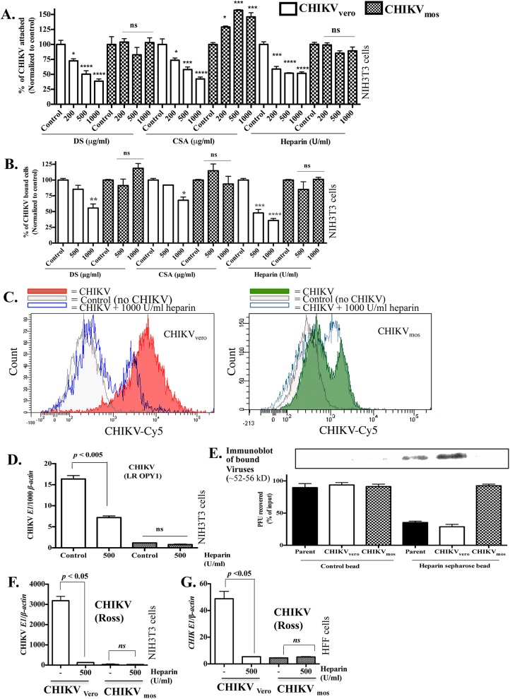
Fig 6. CHIKVvero, but not CHIKVmos, binds to cell surface glycosaminoglycan receptors.
(A) Equal PFUs of CHIKVvero or CHIKVmos (Ross strain) were pre-incubated with the indicated soluble GAGs at 37°C for 1 h. The virus-GAG complexes were added to NIH3T3 cell monolayer (MOI = 1) and incubated at 4°C for 1 h and the GAGs neutralization of viral attachment was measured by RT-qPCR. Data were normalized to controls without GAG treatment. (B) Equal PFUs of CHIKVvero or CHIKVmos (Ross strain) were pre-incubated at 37°C for 1 h with indicated GAGs and added to NIH3T3 cells (MOI = 2.5) at 4°C for 45 min. The virus-bound cells were quantified by flow cytometry and the representative histograms are shown in (C). (D) Heparin (500U/ml) neutralization of CHIKVvero and CHIKVmos (LR-OPY1 strain) attachment was performed in NIH3T3 cells and measured by RT-qPCR. (E) CHIKVvero, CHIKVmos or their parental stock (105 PFU, Ross strain) were mixed with heparin-conjugated sepharose beads or unconjugated sepharose beads (control) at 4°C for 45 min and unbound viruses recovered from beads were quantified by plaque assay in Vero cells (shown in bottom). Input viruses without beads (control) were also quantified by a plaque assay and viruses recovered from the beads were expressed as percentage of the input. Viruses bound to the respective heparin-conjugated and control beads were lysed and also analyzed by immunoblotting assays (shown in top). The effect of heparin pre-treatment (1000 U/ml) on CHIKVvero and CHIKVmos (Ross strain) replication was measured in NIH3T3 (F) and HFF (G) cells at 24 h by RT-qPCR. GAG treated samples were normalized to their respective controls and analyzed using a one-way ANOVA (**** denotes p < 0.0001, *** denotes p < 0.0005, ** denotes p < 0.005, * denotes p < 0.05, and ns denotes p ≥ 0.05). DS, dermatan sulfate; CSA, chondroitin sulfate A.

