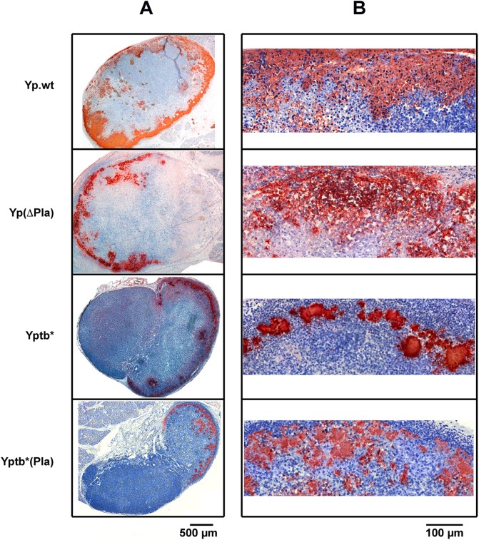
Fig 1. Distribution and infiltration pattern of bacteria in dLNs infected with Y. pestis or Y. pseudotuberculosis strains containing or not the pPla plasmid.
Mice were infected id in the ear with 5x10³ cfu of each strain. At 48 h post infection mice were sacrificed and the ipsilateral superficial parotid LN was collected. LN sections were immunostained with an anti-Y. pestis (first two rows) or -Y. pseudotuberculosis (last two rows) antiserum. Bacteria have an orange-brown coloration, and the tissues are counterstained with hematoxylin (blue). (A) and (B) panels represent low- and high-magnification pictures, as indicated by the corresponding scale bars. Strains are denoted as in Table 1. The figure shows typical aspects observed in a panel of 68 examined LNs (20 infected with Yp.wt, 26 with Yp(ΔPla), 11 with Yptb* and 11 with Yptb*(Pla)).

