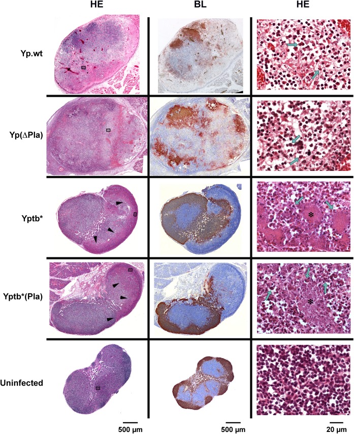
Fig 2. Histology of dLNs infected with Y. pestis or Y. pseudotuberculosis strains containing or not the pPla plasmid.
Mice were infected id in the ear with 5x10³ cfu of each strain and dLNs were collected 48h later. An uninfected LN is shown for comparison. HE: hematoxylin-eosin staining; BL: immunostaining of B lymphocytes (brown), and counterstaining with hematoxylin (blue). In Yp.wt- and Yp(ΔPla)-infected nodes (first two rows): areas of lymphoid tissue depletion and necrosis appear on low-magnification HE pictures (left column) as zones of lower staining intensity or as irregular pink areas; at higher magnification (right column), dissociation of the lymphoid tissue, deposition of eosinophilic fibrillar material, and the presence of necrotic cells and cell debris characterize the necrotic process; typical PMNs harboring horseshoe-shape nuclei are visible (green arrows). In Yptb* and Yptb*(Pla)-infected nodes (third and fourth rows): on low-magnification HE pictures, arrowheads indicate the limits of the inflammatory infiltration clearly delineated from the rest of the organ; higher magnifications show that the inflammatory infiltration is essentially made of PMNs (green arrows), which come in close contact with the bacterial foci (stars). In uninfected LNs (last row), the lymphoid tissue density and follicular organization (B cells at the periphery) are normal. Strains are denoted as in Table 1. Sections of infected dLNs shown here are from the same dLNs as in Fig 1. The position of the fields shown in the right column is marked by grey rectangles in the corresponding low-magnification HE sections.

