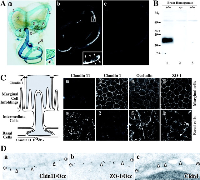
Figure 1.
Localization of TJ proteins in the inner ear of rodents. A, Claudin 11 gene expression is widespread in mouse inner ear. a, X-gal histochemistry reveals expression of lacZ regulated by the endogenous Claudin 11 promoter. 1, Semicircular canals; 2, ampulla; 3, utricle and saccule; 4, suprastrial zone; 5, lateral wall of the cochlear duct; 6, CNS myelin in the eighth cranial nerve. Inset, Strial capillaries (arrow) express Claudin 11. b, Paraffin section of wild-type cochlea labeled with anti-claudin 11 antibodies (37E3) shows expression in the stria vascularis. Inset, Antibodies label the basal cell layer (arrows) but not the marginal cell layer (arrowheads). c, Secondary antibodies do not label a section adjacent to that in b. B, Specificity of anti-claudin 11 antibodies. Antibodies used in Ab label a 24 kDa band on a Western blot of whole brain from wild-type (+/+, lane 1) but not Claudin 11-null (-/-, lane 2) mice. Secondary antibodies do not label the Western blot (+/+, lane 3). Mr, relative mobility (in kilodaltons). C, Claudin 11 expression in rat stria vascularis. The schematic shows intimate relationships between different cells in the stria vascularis. a-h, Confocal micrographs reveal labeling of strial marginal cells (a-d) and basal cells (e-h) using antibodies against TJ proteins claudins 11 and 1, occludin, and ZO-1. D, Immunoelectron micrographs of TJ proteins in basal cells. Colocalization of claudin 11 and occludin (a) ZO-1 and occludin (b) but not claudin 1 (c) at membrane “kiss” sites in basal cell TJs is shown. Cldn 11, claudin 11; Occ, occludin; Cldn 1, claudin 1.

