
Figure 6.
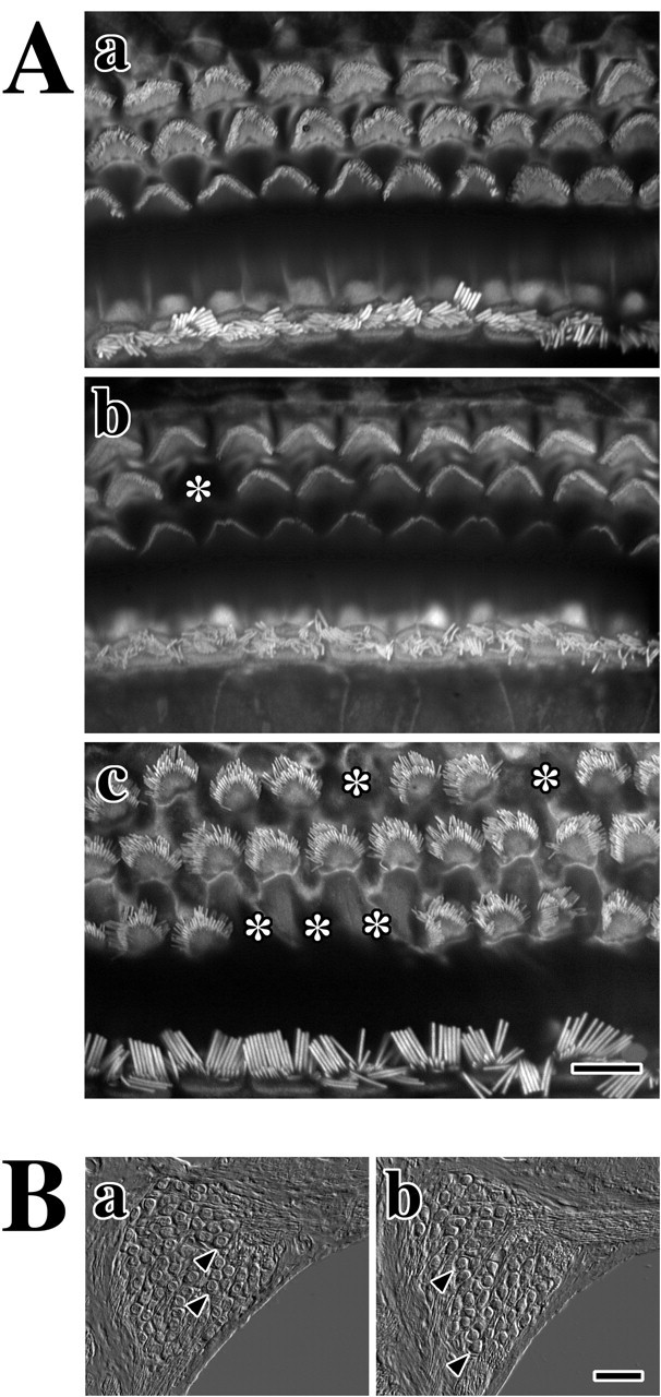
Normal morphologies of hair cells and spiral ganglion neurons in adult Claudin 11-null mice. A, Fluorescence microscopy from phalloidin-labeled hair cell stereocilia in organ of Corti from adult mice. a, Outer and inner hair cells from the basal cochlear turn of a P90 wild-type mouse. b, c, Outer and inner hair cells from the basal cochlear turn (b) and the apical turn (c) of a P90 Claudin 11-null mouse. Asterisks mark sites of individual OHC loss. Scale bar, 10 μm. B, Differential interference contrast microscopy showing cross sections of spiral ganglia in the midcochlear region reveal neuron cell bodies (arrowheads) from P90 wild-type (a) and Claudin 11-null (b) mice. Scale bar, 40 μm.