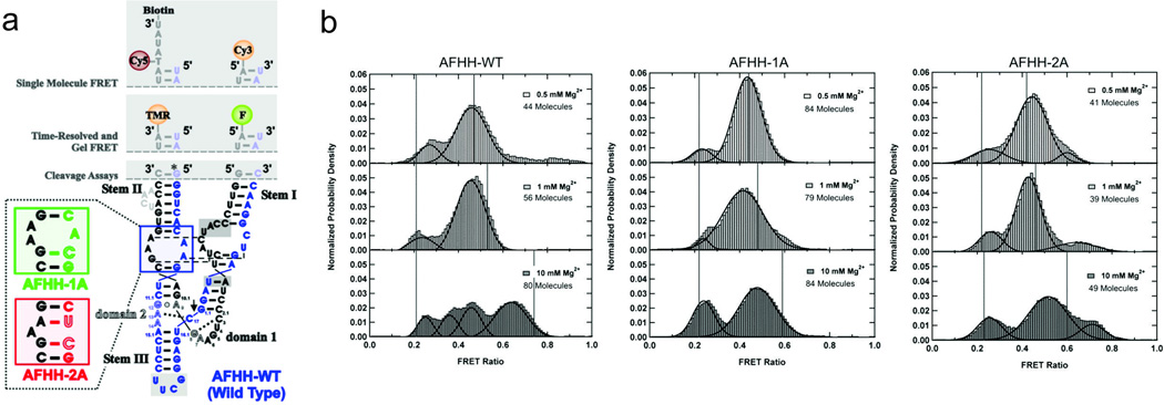
Fig. 3.

Single-molecule FRET investigation of the hammerhead ribozyme. a Full-length hammerhead ribozyme used in the studies discussed. The labeling sites for single-mol ecule FRET are indicated at the top, and the mutations used to disrupt loop-loop interactions are shown in the bottom-left. b Single-molecule FRET histograms showing the effect of magnesium concentration and loop mutations on the conformations adopted by wild-type (left panel) and two mutant (center and right panels) ribozymes. The active, high-FRET conformation is sampled only by the wild-type ribozyme, and only under high magnesium conditions (McDowell et al. 2010)
