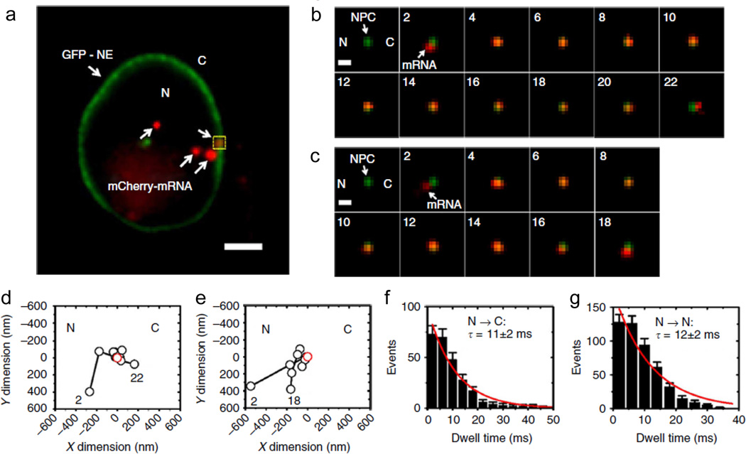
Fig. 7.
Tracking single mRNPs through the nuclear pore complex in HeLa cells using SPEED microscopy. a Wide-field epifluorescence illumination of four mCherry-tagged mRNPs (red) and GFP-nuclear envelope (green). b A successful mRNP export event through the NPC captured using SPEED microscopy. c An abortive mRNP export event. d, e Single particle tracks (black) and the centroid of the NPC (red) of successful and abortive mRNP export events through the NPC, as shown in b and c, respectively. f, g Export time distributions and single-exponential fit of successful (f) and abortive (g) mRNP export events through the NPC as shown in b and c, respectively (Ma et al. 2013)

