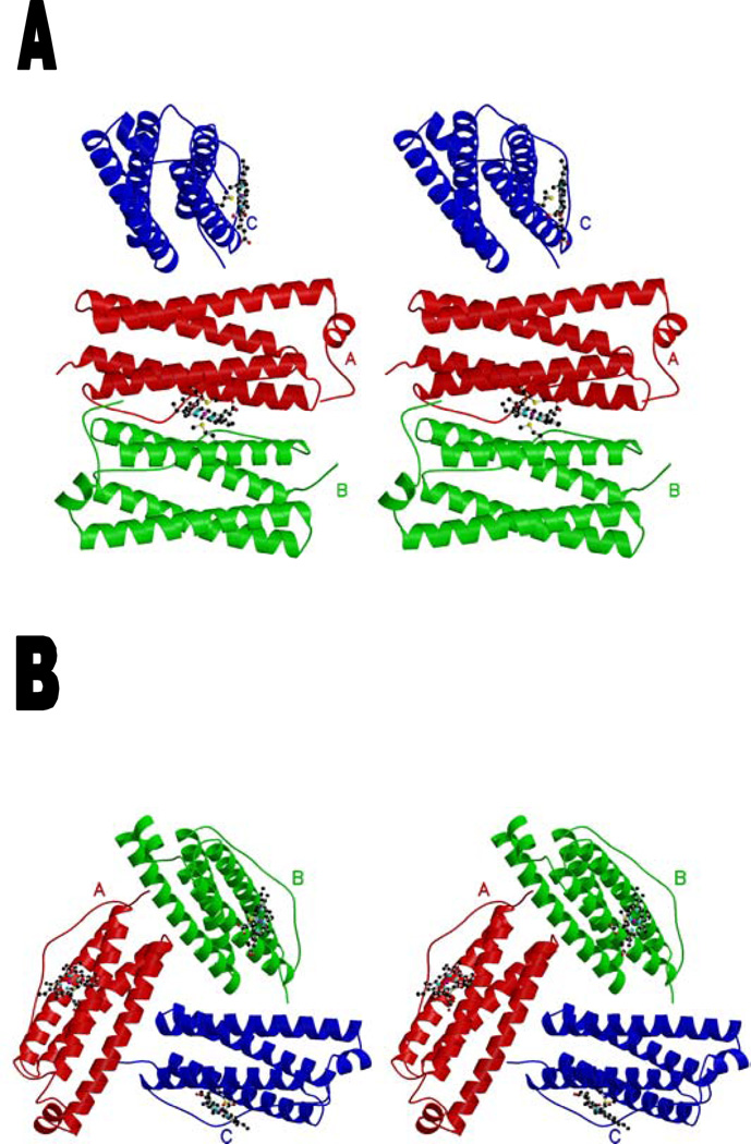
Figure 1.
stereoview of the Rhodobacter capsulatus bacterioferritin subunits of the asymmetric unit schematized as a ribbon. The heme group is represented in ball-and-stick. Monomers related by a noncrystallographic 2-fold axis are colored in red (A) and green (B). The subunit (C) binding to the heme situated on the 2-fold crystallographic axis is colored in blue. (a) Trimeric structure including one heme with its enclosing noncrystallographic dimer and one heme on a crystallographic axis (at occupancy 0.5) with one monomer of its enclosing dimer. (b) Trimeric structure with proper 3-fold symmetry, including three heme moieties at 0.5 occupancy. The Met52 and heme are represented in ball-and-stick. The drawing was generated using Molscript (19) and Raster3D (20).

