Figure 1.
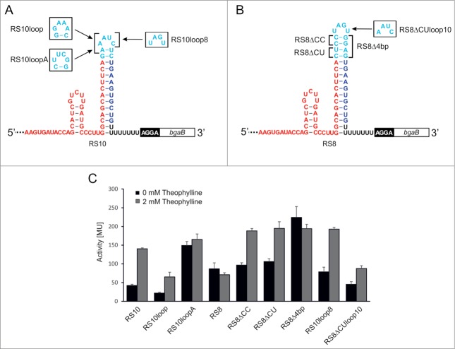
Riboswitch variants and activity tests. Sequence and secondary structure of the functional riboswitch 10 (RS10), the non-functional riboswitch 8 (RS8) and tested mutants are shown in (A) and (B), respectively. Shine-Dalgarno sequence (black box) and bgaB reporter gene (white box) follow immediately downstream of the riboswitches. Aptamer sequence is shown in red, the spacer region in cyan, and the sequence complementary to the aptamer is indicated in blue, followed by a black U-stretch. These elements make up the intrinsic terminator element. Mutated positions are indicated. (C) Activity test of β-galactosidase reporter constructs with RS10 and RS8 mutants. Activities are indicated in Miller Units. The reduction in activity observed for RS8 and RS8Δ4bp is statistically not significant according to Student's t-test (p = 0.17).

