Figure 2.

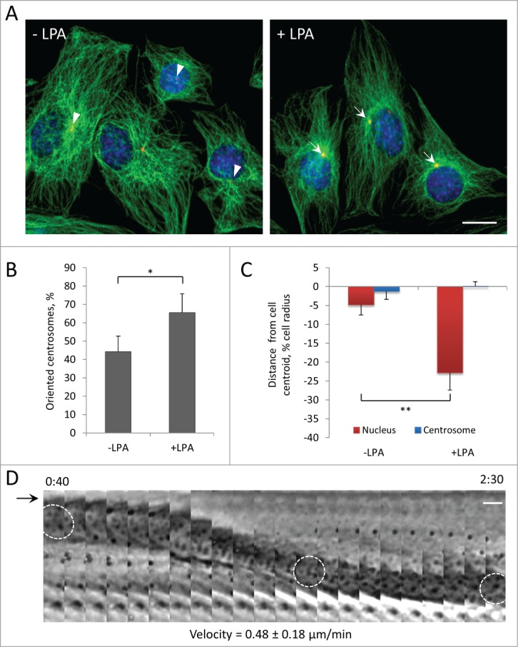
Centrosome orientation in wounded monolayers of serum-starved C2C12 myoblasts is stimulated by LPA and occurs by rearward movement of the nucleus. A, Immunofluorescence images of microtubules (green), pericentrin (red) and nuclei (blue) in C2C12 myoblasts at the edge of a wounded monolayer before and after 10 μM LPA stimulation for 2 hr. Arrows indicated oriented centrosomes; arrowheads indicate unoriented centrosomes. B, Quantification of centrosome orientation in C2C12 myoblasts before and after LPA stimulation. Centrosomes were scored “oriented” if they were within the pie-shaped region defined by the nucleus and the 2 sides of the leading edge; random orientation is 33% by this measure.36 C, Positions of the nucleus and the centrosome along the front-back cell axis for the cells treated as B. The cell centroid is defined as “0;" + values, toward the leading edge; - values, toward the cell rear. D, Kymograph showing the rearward movement of the nucleus (outlined in 3 panels) from a phase contrast movie of a wound edge C2C12 myoblast stimulated with LPA. The arrow denotes the leading edge of the cell, which does not extend with LPA treatment.2 Time is in hr:min after LPA treatment. The velocity of nuclear movement from 20 cells is indicated. Bars in A and D, 10 μm. Error bars in B and C are SD from 3 experiments (n > 30 cells per experiment). *, P < 0 .05; **, P < 0 .01 by t-test.
