Figure 1.

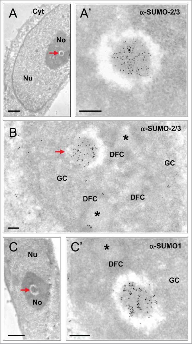
Characterization of INBs by I-EM localization of SUMO-2/3 and SUMO1 (ultra-thin sections of glutaraldehyde-fixed, Lowicryl embedded HeLa cells). (A) The INB (arrow) is electron-dense and centrally-located. (A’) Enlargement showing high SUMO-2/3 content as detected with primary anti-SUMO-2/3 antibody and a secondary antibody conjugated to 5 nm gold particles. Nu = Nucleus, No = Nucleolus, Cyt = cytoplasm. Scale bars: 1 and 0.2 µm in A and A’, respectively. (B) Compared to the classical nucleolar components, the INB (arrow) is surrounded by a white halo and highly labeled by the anti-SUMO-2/3 antibody (10 nm gold particles) whereas the FCs (asterisks) surrounded by the DFC and embedded within the GC are unlabelled. Scale bar = 0.2 µm (C–C’) As in A, except that the centrally-located INB (arrow) is labeled, as shown in C’, with an anti-SUMO1 antibody and secondary antibody conjugated to 10 nm gold particles. Scale bars: 1 and 0.2 µm, respectively.
