Figure 2.

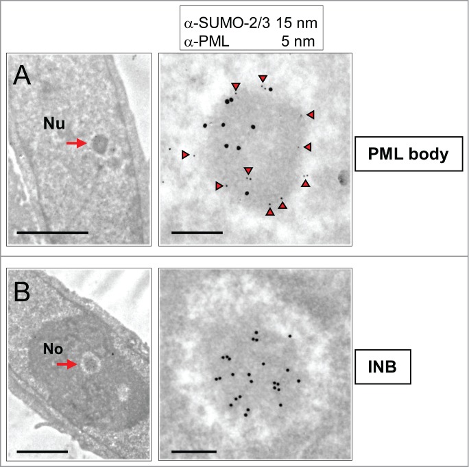
INBs are distinct from PML bodies as shown by differential PML and SUMO-2/3 content. (A) A nucleoplasmic NB (arrow, left frame), double-labeled (as shown in enlarged right frame) with mouse anti-PML (5 nm gold particles underlined by arrow-heads) and rabbit anti-SUMO-2/3 (15 nm gold particles) antibodies is identified as a PML body. Notice peripheral PML-labeling. Scale bars: 2 and 0.2 µm, respectively. (B) On the same thin-section, a centrally-located NB (arrow, left frame) labeled by anti-SUMO-2/3 (15 nm gold particles, right frame) but not anti-PML (lack of 5 nm gold particles) antibodies is defined as an INB. Nu: nucleoplasm, No: Nucleolus. Scale bars: 2 and 0.2 µm, respectively.
