Figure 4.

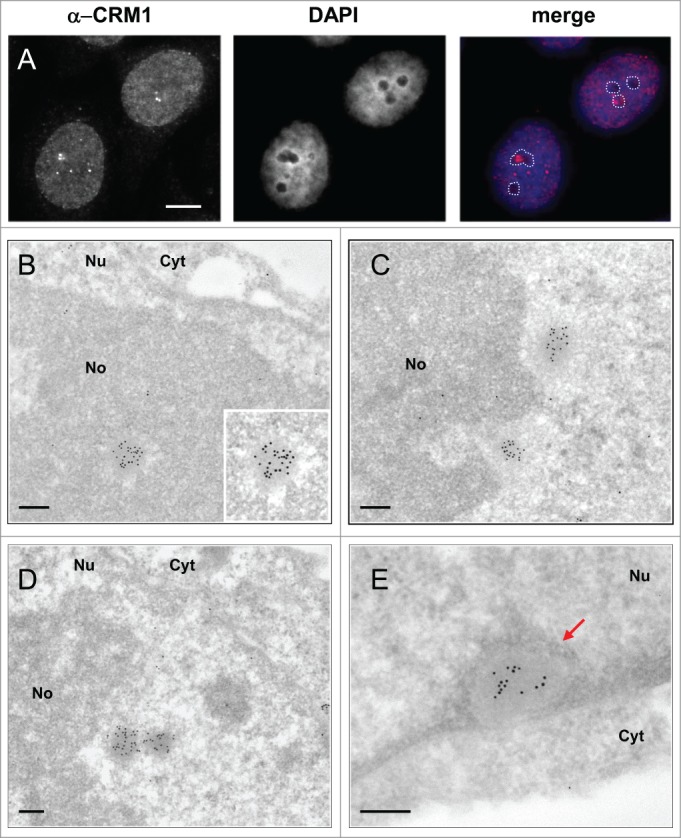
Ultrastructural characterization and nuclear distribution of CNoBs. (A) IF localization of CNoBs in HeLa cells. Nucleolar association is detected by merging CRM1 foci with nucleoli (unstained with DAPI, dotted lines). CNoBs associated with - and distant from - nucleoli are evidenced. Scale bar: 5 µm. (B–D) I-EM detection of CRM1 (glutaraldehyde-fixed, lowicryl-embedded HeLa cells, 10 nm gold particles) reveals uniform, vitreous aspect of CNoBs within the nucleolus in B, at the nucleolar periphery in C and D or within the nucleoplasm as in D, (right object) and E. CNoB in (E), in close proximity to the nuclear envelope, is surrounded by peripheral nuclear heterochromatin (arrow). Nu: nucleus, No: nucleolus, Cyt: cytoplasm. Scale bars: 0.2 µm.
