Figure 6.

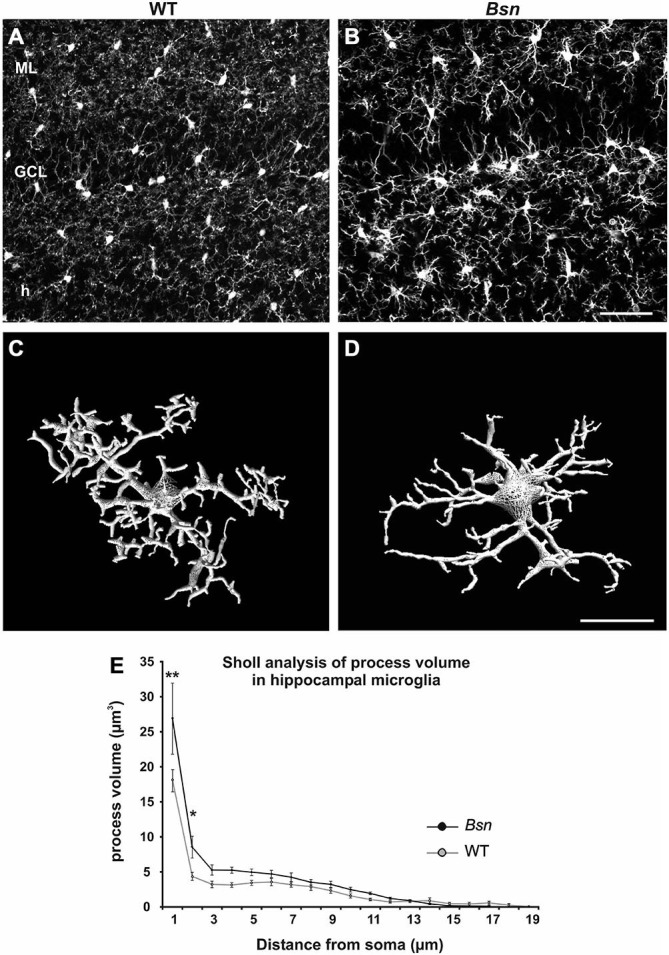
Hippocampal microglia from Bsn-mutant mice show altered morphology. Comparative Iba1 staining in hippocampi from WT and Bsn-mutant (A) In WT mice, microglia are evenly distributed throughout the layers of the DG and are characterized by a relatively small, round soma with thin, finely-branched processes. (B) A similar distribution was observed in the Bsn DG, however, microglia exhibited larger somata and thicker processes. (C,D) 3D-reconstruction of Iba-1-labeled microglia in the hilus depicts characteristic morphological changes at the cellular level. (C) Microglia from WT animals displayed ramified processes that were radially distributed around the cell soma. (D) Microglia from Bsn-mutants showed enlarged cell bodies and thicker processes close to the soma. (E) Sholl analysis confirmed the latter finding. Data are expressed as mean ± SEM. *p < 0.05; **p < 0.01. Scale bars: A,B = 50 μm; C,D = 2 μm. Abbreviations: ML, molecular layer; GCL, granule cell layer; h, hilus.
