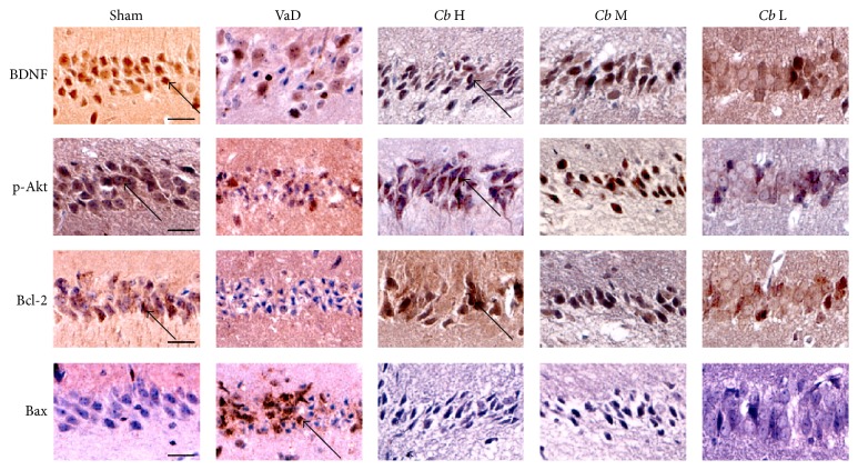
Figure 6.
Immunohistochemical staining. Representative images of the immunohistochemical staining of BDNF, p-Akt, Bcl-2, and Bax in area CA1 of the hippocampus. Cells with a brown-stained cytoplasm are considered positive. Sham, sham-operated group; VaD, VaD model group; Cb H, C. butyricum- (1 × 10⁸ CFU-) treated group; Cb M, C. butyricum- (1 × 10⁷ CFU-) treated group; and Cb L, C. butyricum- (1 × 10⁶ CFU-) treated group. Magnification: 400x. Scale bar = 20 μm.

