Figure 6.

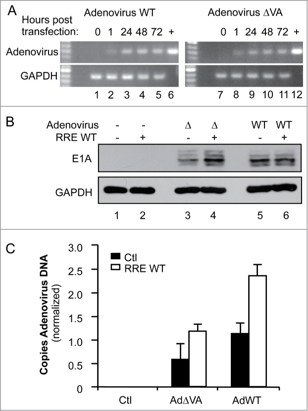
RRE restores the replication of VA-defective adenovirus. (A) Amplification of adenovirus DNA. HeLa cells were transfected with 1 μg of pcDNA3 (lanes 1, 7), pAd5 WT (lanes 2–5), or pAd5ΔVA (lanes 8–11). DNA was collected at 1 h (lanes 2, 8), 24 h (lanes 3, 9), 48 h (lanes 1, 4, 7, 10) or 72 h (lanes 5, 11) post transfection. (+) represents amplified plasmid DNA. Viral and GAPDH DNAs were amplified by 25 and 30 cycles of PCR, respectively. (B and C) HeLa cells were cotransfected with 1 μg of pcDNA3 (lanes 1–2), pAd5ΔVA (lanes 3–4), or pAd5 WT (lanes 5–6), and with 1μg of pcDNA3 (lanes 1, 3, 5) or pcDNA3-RRE (lanes 2, 4, 6). (B) Expression of adenoviral proteins. Expression of adenoviral proteins was assessed by immunoblot with anti-E1A protein compared to the total proteins represented by GAPDH expression. (C) Quantitation of adenoviral DNA. Viral DNA was quantified by real-time qPCR using a FAM-TAMRA probe. The histogram represents the average of 3 independent experiments ± SEM.
