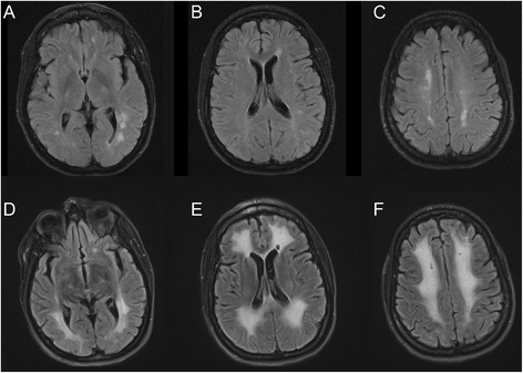
Fig. 2.

Case 2: MR images (FLAIR) of brain lesions. a-c On admission foci of increased signal are present in the white matter of the cerebral hemispheres bilaterally, predominantly in a periventricular distribution. d-f Repeat imaging seven weeks later, after further neurological deterioration, shows prominent interval progression of signal abnormality in the subcortical and periventricular white matter
