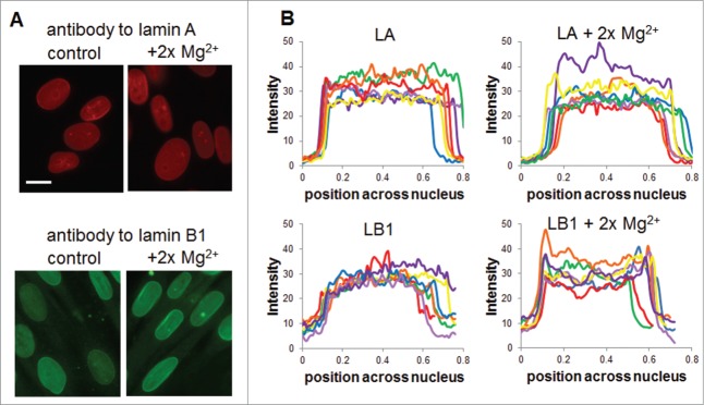
Figure 6.

Nucleoskeleton redistribution with divalent cations. Human foreskin fibroblasts grown in standard cell culture media or in media spiked with 2x Mg2+ were fixed and labeled with antibodies for lamin A and lamin B1. (A) Representative image of each group shows mild changes in lamin A intensity or localization in the presence of Mg2+ whereas lamin B1 shows altered localization to the nuclear periphery. (B) Quantification of localization in nuclei from more than 10 fields of view each using the scheme shown in Figure S1 shows altered lamin B localization. Scale bar is 20 μm.
