
Figure 1.
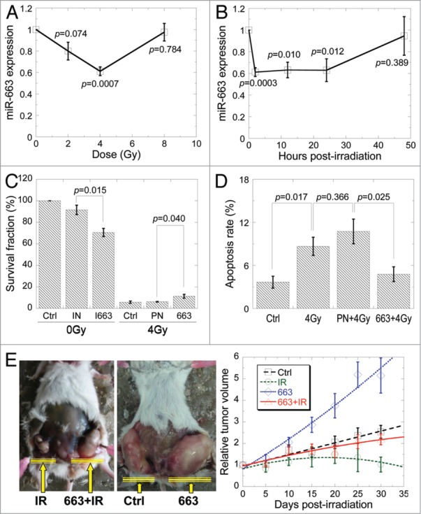
MiR-663 was downregulated in directly irradiated cells. (A) qRT-PCR was performed to detect miR-663 expression in HeLa cells 2 h after exposure to different doses of X-rays. (B) MiR-663 levels in HeLa cells exposed to 4 Gy X-ray irradiation at different time-points were detected using qRT-PCR. (C) Colony forming assay was performed on HeLa cells subjected to different treatments, after which cells were allowed to grow for 13 d. 663, miR-663 mimics; I663, miR-663 inhibitors; PN, negative control for miR-663 mimics; IN, negative control for miR-663 inhibitors. (D) Apoptosis rates of HeLa cells subjected to 4 Gy X-ray irradiation plus transfection with either miR-663 mimics or negative control (PN) were assayed with Hoechst33342/PI double staining. (E) Tumors generated by a stable HeLa cell line with inducible miR-663 expression in NOD/SCID mice 1 mo after 5 Gy X-ray irradiation and/or the induction of miR-663 expression. Tumor volumes were measured every 5 d and normalized to those obtained immediately before irradiation. The tumor volumes were measured using the formula ab2 × π/6, whereby a represents the length and b is the width. n = 10 flanks. Data were obtained from at least three independent experiments, and presented as means ± SE P values between the indicated samples and sham control (Ctrl) are additionally presented.