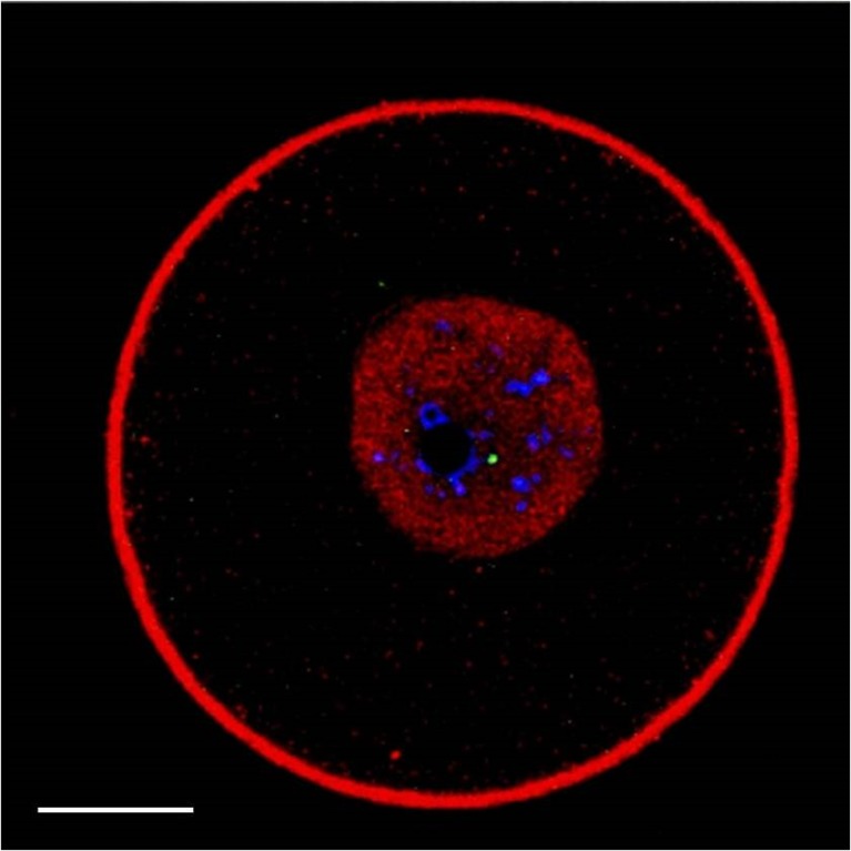
Fig. 3.
Representative image of a GV-stage oocyte stained for γH2AX (green), chromatin (blue), actin (red) and Rad51 (red). In addition to showing a site of damaged DNA and the chromatin organised around the nucleolus or partly dispersed, the nucleus displays an intense and homogeneous red staining. Under the conditions applied in this study, actin is never observed in the GV (see also the “Results” section). Therefore, intranuclear red staining corresponds to Rad51 localisation. Scale bar equals 15 μm

