
Fig. 2.
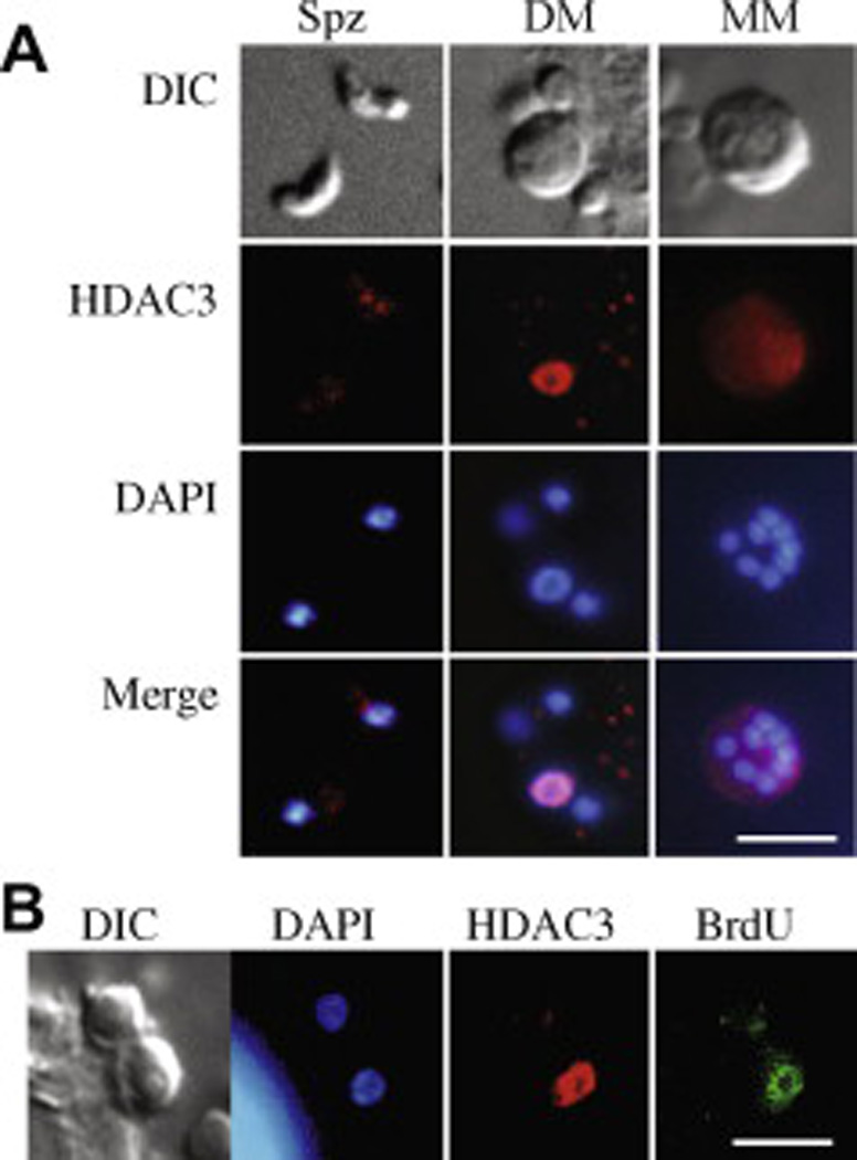
Immunolocalisation of Cryptosporidium parvum histone deacetylase 3 (CpHDAC3). (A) Different life stages include sporozoites (Spz), developing meronts (DM) and a mature type I meront (MM). Developing meronts whose nucleus adopted a toroidal appearance expressed CpHDAC3 at high levels and this signal overlapped that of the DAPI stain (evident in the merged images). (B) Immunolocalisation of recently incorporated bromodeoxyuridine (BrdU, green) indicated that DNA replication occurred in the meront that displayed high levels of CpHDAC3 but not the adjacent meront. The bright signal in the lower left corner of the DAPI panel was from a human host cell. DIC = differential interference contrast. The white bars represent 5 µm.