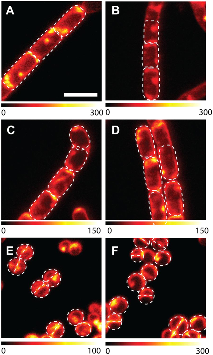
Figure 3. Localization of Hyp-apoMb in different types of bacteria.
Selected STED images of B. subtilis (A,B), E. coli (C,D) and S. aureus cells (E,F) incubated with Hyp-apoMb (10 μM Hyp, 30 μM apoMb) collected under excitation at 566 nm and detection at 605/70 nm. The STED beam was at 715 nm, power 30 mW and dwell time 0.1 ms (A–D) and 0.05 ms (E,F). White dashed lines are intended as visual aid to guide the eye along the bacterial shape. Scale bar, 2 μm.

