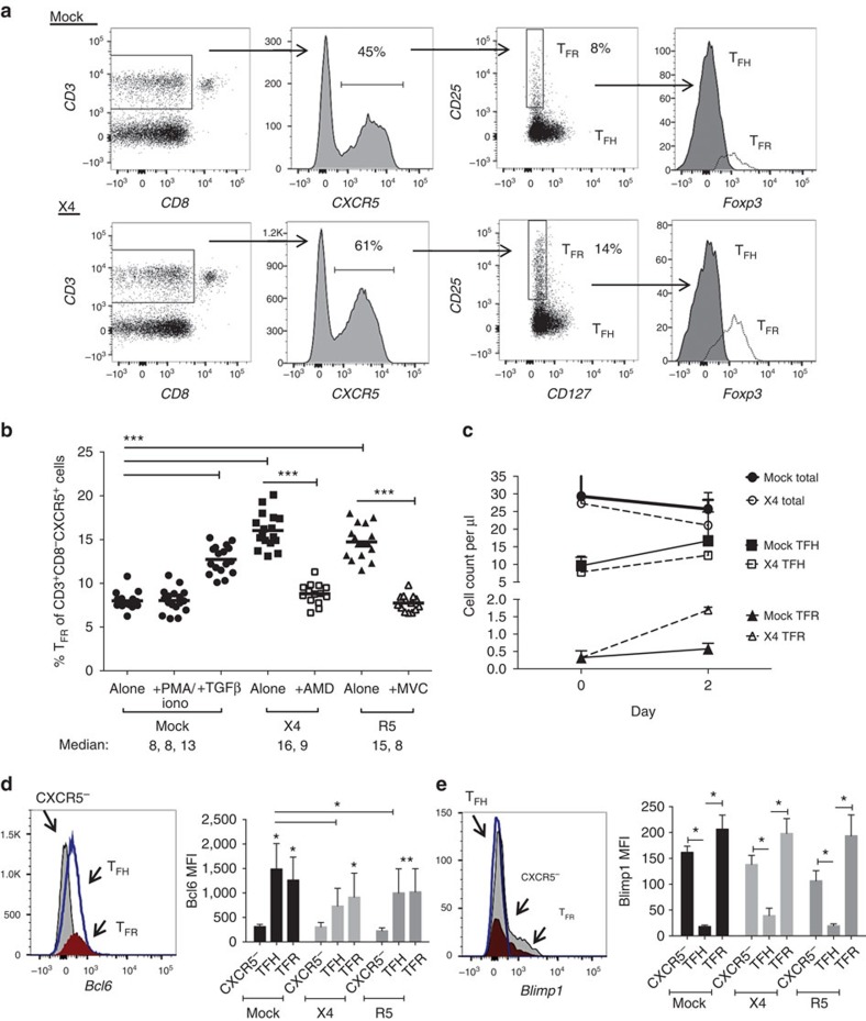
Figure 3. HIV entry and replication promote TFR expansion.
Disaggregated tonsil cells were spinoculated with X4 or R5 HIV and TFR populations were analysed by flow cytometry (n=15). (a) A representative example of tonsil cell flow gating. From viable CD3+CD8− cells, TFR are defined as CXCR5+ and CD25hiCD127–. TFR cells contain Foxp3+ cells, whereas remaining TFH (CXCR5+CD25lo/−) cells are Foxp3−. (b) Percentages of TFR determined by gating strategies in a are shown. Experimental conditions include mock-spinoculated cells cultured with PMA (50 ng ml−1) and ionomycin (1 μg ml−1) or exogenous TGF-β (100 ng ml−1) for 24 h and cells pretreated to block CXCR4 (AMD, 200 μM) and CCR5 (MVC, 2 μM). (c) Using flow cytometry counting beads, the number of cells per μl were determined for total (CD3+CD8−), TFH (CXCR5+CD25lo/−) and TFR (CXCR5+CD25hiCD127−) subsets in mock- and X4-spinoculated samples (n=3). (d) Bcl-6 expression is shown in CXCR5− (grey), TFH (blue) and TFR (red) populations after mock-, X4- or R5-spinoculation (n=5). (e) Blimp-1 expression was also determined as in d. The horizontal bars of each graph indicate the median value and are listed where appropriate for clarity. Statistical analyses were performed by Friedman nonparametric tests (b,d,e) and significance is denoted by asterisks where *P<0.05, **P<0.01 and ***P<0.001.

