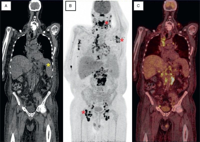
FIGURE 1.

Slices of fluorodeoxyglucose PET combined with computed tomography (A: CT; B: PET; C: fusion of PET/CT). Besides a splenomegaly (yellow asterisk) a very intensive uptake was measured in nearly all lymph node stations. In particular, pathologic [18F]2-fluoro-2-deoxy-D-glucose accumulation was detected in cervical, axillary, mediastinal, paraaortic, and inguinal lymph nodes (red asterisks) as well as in the tongue, thyroid, and lung (MIP–PET). CT = computed tomography, MIP = maximum intensity projection, PET = positron emission tomography.
