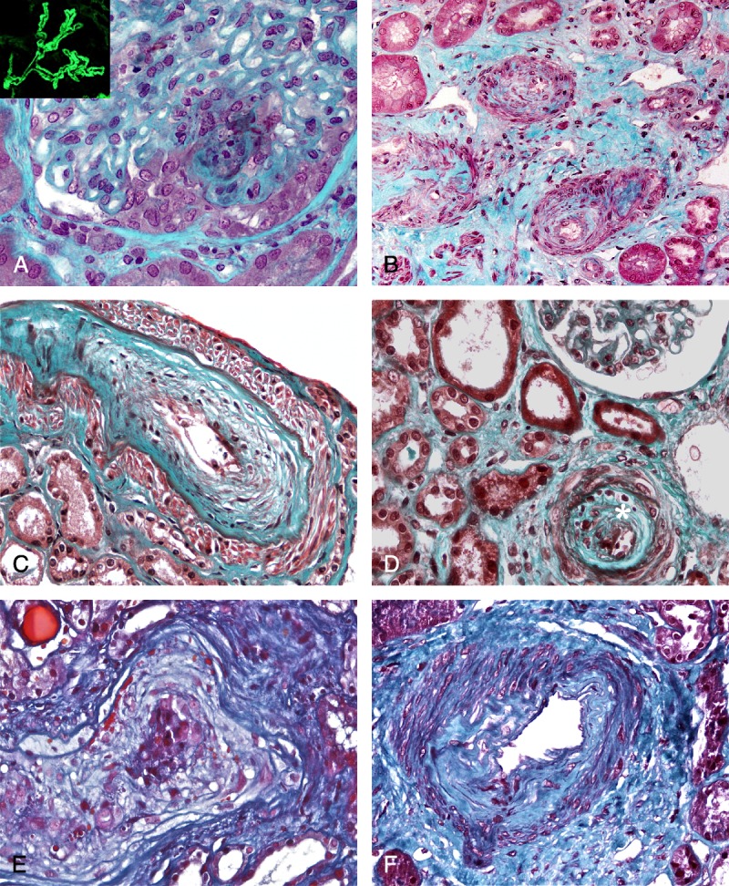
FIGURE 2.

A, Kidney biopsy (light microscopy, Masson trichrome, ×400) performed in a 45-year-old woman (Table 5, Patient 4) with a history of antisynthetase syndrome and MN diagnosed 10 years earlier. She presented with an exacerbation of the antisynthetase syndrome and an AKI. Kidney biopsy disclosed a crescentic MN with extracapillary proliferation noted in 11/20 glomeruli. Test for ANCA was negative. (Insert shows IgG staining.) B, Kidney biopsy (light microscopy, Masson trichrome, ×200) performed in a 68-year-old normotensive man (Table 5, Patient 6) with a history of DM who presented with AKI. Edematous thickening of the intima of interlobular arteries associated with interstitial fibrosis and ischemic glomerular lesions was noted. C and D, Kidney biopsy (light microscopy, Masson trichrome, ×400) performed in a 67-year-old normotensive woman (Table 5, Patient 7) who presented with an exacerbation of PM (rhabdomyolysis, lung involvement) and AKI. Kidney biopsy disclosed edematous thickening of the intima of an interlobular artery superimposed on chronic arteriosclerosis. Mild lymphocytic infiltration (*) was detected in 2 interlobular arteries within the marked fibrous intimal thickening (2D). E, Kidney biopsy (light microscopy, Masson trichrome, ×400) performed in a 55-year-old normotensive man (Table 5, Patient 5) who presented with PM (rhabdomyolysis, interstitial pneumopathy) and AKI. Pathologic analysis disclosed the presence of marked edematous thickening of the intima of the interlobular arteries leading to an obstruction of the vessel lumen. F, Kidney biopsy (light microscopy, Masson trichrome, ×400) performed in a 34-year-old normotensive man (Table 5, Patient 8) who presented with DM (rhabdomyolysis, interstitial pneumopathy), mild proteinuria (0.8 g/d and subsequently 1.2 g/d), and hematuria. On pathologic examination, a marked fibrous thickening of the intima of an arcuate artery was present.
