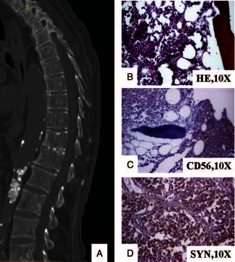
FIGURE 3.

The vertebral metastases are seen as multiple osteoblastic-osteolytic foci at CT-scan (A). Under microscope, small cells can be observed in the bone marrow (B) that are marked by CD56 (C) and synaptophysin (D). CD = cluster of differentiation, CT = computer-tomography.
