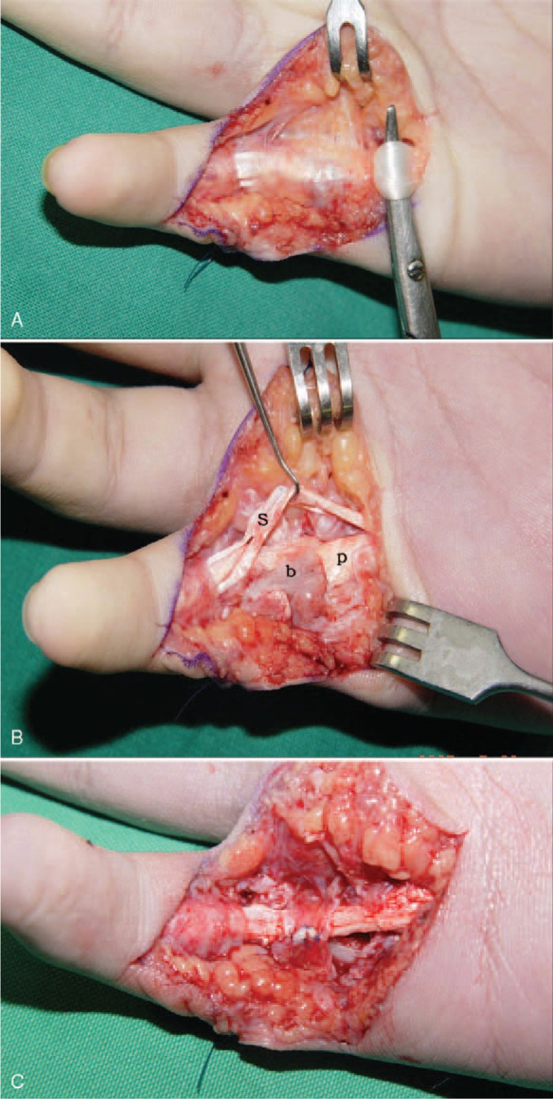
FIGURE 3.

A–C. Intraoperative photographs show: (A) Flexor tendons adhesion at the fracture site. (B) FDP is interposed between the epiphysis and volarly displaced metaphysis and fracture is united. (b: bone, p: FDP, s: FDS) (C) A2 pulley is repaired after volarly displaced bone had been removed and FDP has been freed.
