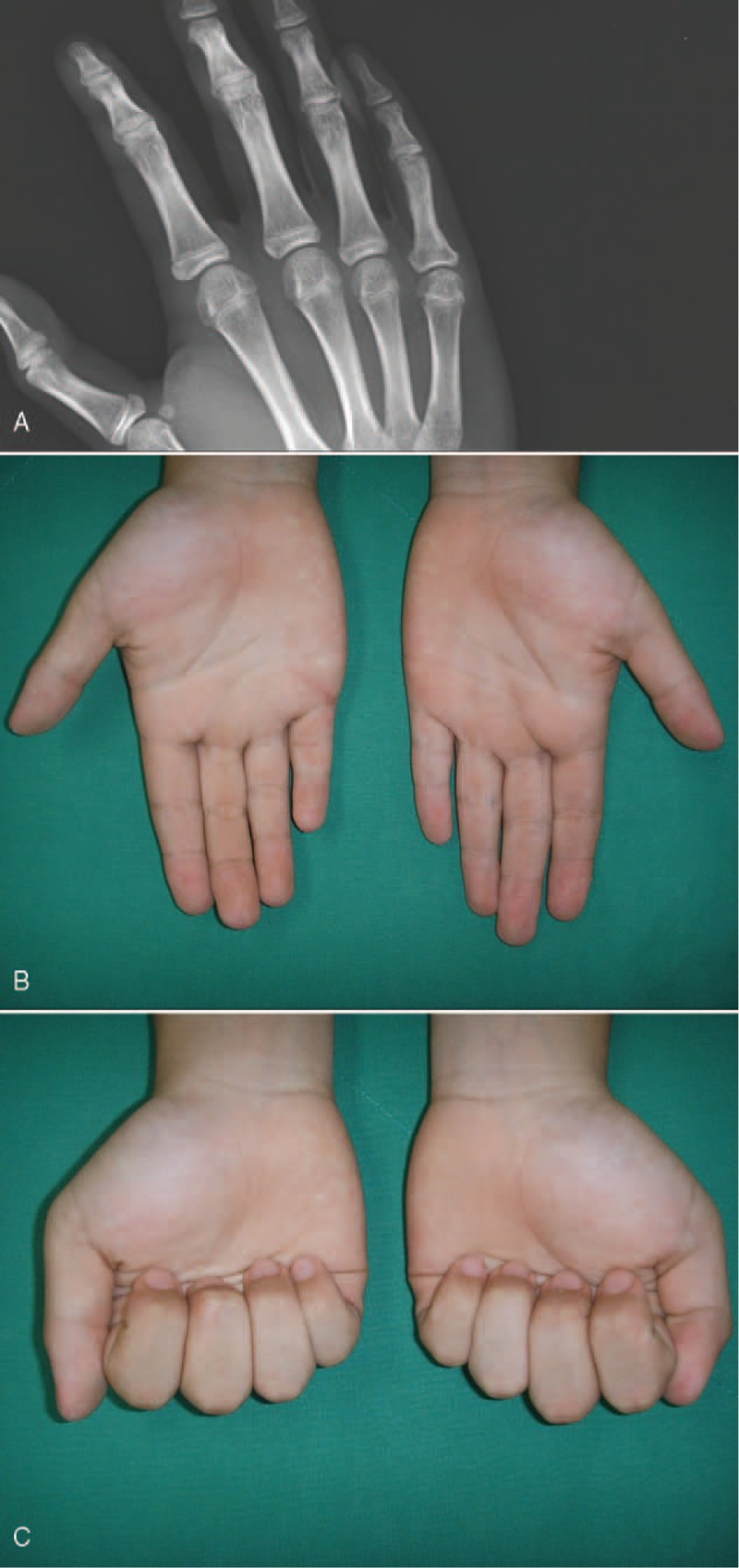
FIGURE 5.

AC (A) An oblique radiograph obtained 12 months after operation shows the growth plate of proximal phalanx of small finger closed naturally. (B, C) Photographs obtained 12 months after operation show a normal range of motion and alignment of a right small finger.
