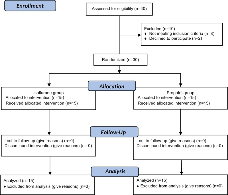
FIGURE 1.

Ophthalmologic, urologic, gastroenterological, tilt table, and MRI imaging. A: Anisocoria right < left before (left image) and 1 hour after 5% cocaine-HCl (right image); B: videourodynamics: detrusor hypocontractility and urinary retention at 15 (left: 600 mL) and 60 minutes (right: 800 mL) after micturition; C: colonic pseudoobstruction 2 (left) and 8 hours (right) after barium meal; D: tilt-table testing with abnormal rise in heart rate (fat line +44 bpm) before (left) and with normal heart rate increase (+16 bpm) after IvIg therapy (right). Systolic and diastolic pressures are shown by thin lines. Vertical lines are marking the period of upright tilt. E: Normal spinal MRI scan, thoracolumbar (left) and cervicothoracal (right); F: Normal pontine plane of cranial MRI scan.
