
FIGURE 1.
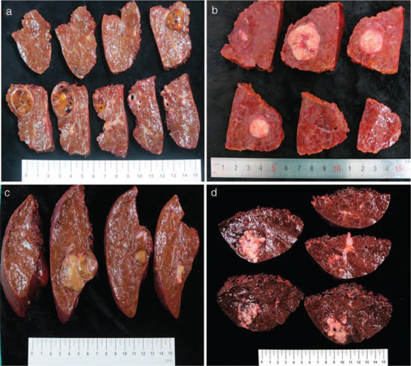
Photographs of hepatocellular carcinoma with different morphological appearance. (A) Type 1, an oval nodule with clear boundary as well as an obvious fibrous capsule; (B) type 2, a roughly oval nodule with a local protrusion at the bottom (arrow); (C) type 3, a lobulated lesion with 2 nodules merging together, each nodule with a clear margin; (D) type 4, a rather irregular lesion with multiple invasion to liver parenchyma.