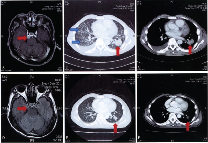
FIGURE 3.

Meningeal metastases in the cerebropontin area in case C (panel 3A). Chest CT revealed a tumor located in the left lower lung and multiple interstitial infiltrations (panels 3B and 3C). Chest CT showed a partial response after afatinib treatment for 7 months. Brain MRI found no evidence of recurrence following afatinib treatment for 9 months. CT, computed tomography; MRI, magnetic resonance imaging.
