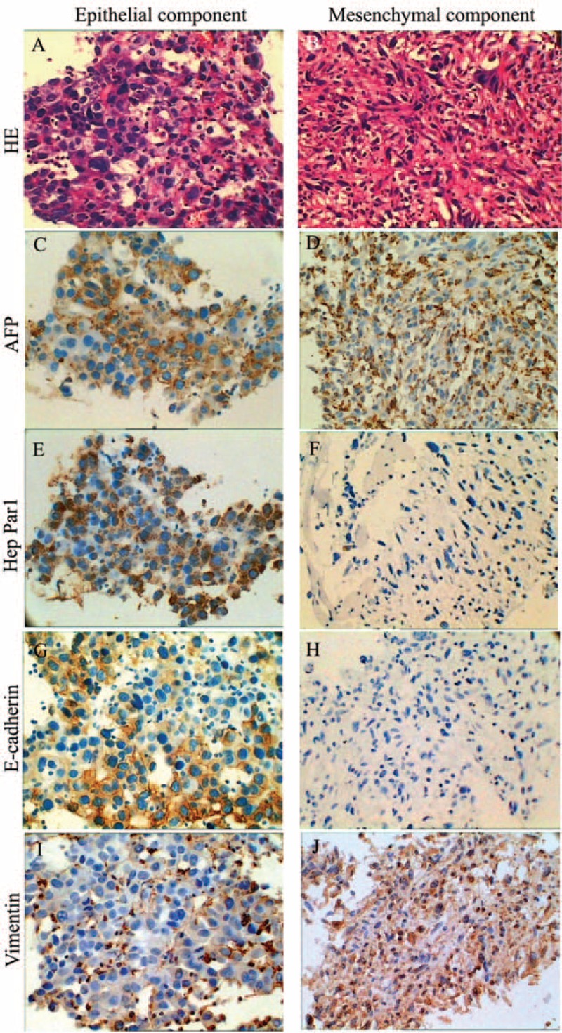
FIGURE 2.

Pathological and immunohistochemical analysis of tumor biopsy from 2 segments. (A) Biopsy revealed poorly differentiated HCC in segment 7. (B) Spindle-shaped cells consisted of a mesenchymal component in the tumor in segment 6. (C)–(J) Tumor cells in both sections were diffusely positive for AFP (C and D). The epithelial component was focally positive for Hep Par1 (E), E-cadherin (G), and vimentin (I). The mesenchymal section was negative for Hep Par1 (F) and E-cadherin (H), and diffusely positive for vimentin (J). AFP = serum a-fetoprotein, HCC = hepatocellular carcinoma.
