
FIGURE 2.
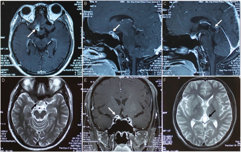
Radiological evaluation of the brain before surgery. T1-weighted axial gadolinium-enhanced MRI demonstrated a ring-enhancing cystic lesion at the right part of suprasellar region in the anterior of optic chiasma (arrows) (A, C, D). T2-weighted image demonstrates the same lesion as in the previous image (arrow) (B). A cyst in the pineal region was also showed with long-T1 and long-T2 signals (arrows) (E, F).