Fig. 1.
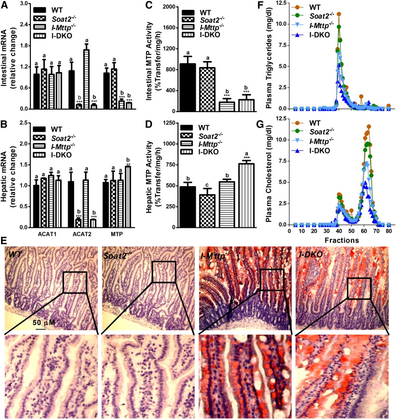
Effect of global ACAT2 and intestine-specific MTP deficiency on intestinal gene expression, lipid accumulation, and plasma lipoproteins. A–D: Total RNA isolated from the intestine (A) and the liver (B) of 12-week-old WT, Soat2−/−, I-Mttp−/−, and I-DKO (n = 5) male mice fed a chow diet was used to quantify mRNA levels of ACAT1, ACAT2, and MTP. Intestinal (C) and hepatic (D) tissues were also used to measure MTP activity. Data are presented as mean ± SD. **P < 0.01 and ***P < 0.001 compared with WT as determined by Student’s t-test. Statistically significant differences in different parameters in the four groups were evaluated by one-way ANOVA with Newman-Keuls multiple comparison test. Different letters above bars for each component indicate statistically significant differences in the mean values in different groups (P < 0.05) as determined by one-way ANOVA. E: Proximal intestinal sections were used for lipid staining by Oil Red O. A higher magnification image of the boxed area is shown under each picture to show the presence of lipids in the absorptive epithelial cells. F, G: Plasma was separated by gel filtration to determine mass of triglycerides (F) and cholesterol (G) in different lipoproteins.

