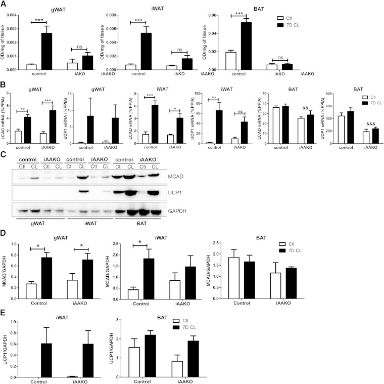
Fig. 7.
Expansion of mitochondrial respiration by β3-AR agonist treatment requires adipocyte ATGL. A: Mitochondrial respiration in adipose tissue depots (n = 4–5) as determined by reduction of the electron acceptor dye TTC. Data were analyzed by two-way ANOVA to determine the effect of CL (*** P < 0.001; ** P < 0.01; * P < 0.05; ns, nonsignificant). B: Adipose tissues from control or iAAKO mice treated with vehicle (Ctl) or CL for 7 days were collected, and the mRNA expression of MCAD and UCP1 was measured by qPCR. C: Western blot analysis and quantification of MCAD (D) and UCP1 (E) protein expression. Data were analyzed by two-way ANOVA to determine the effect of CL (*** P < 0.001; ** P < 0.01; * P < 0.05; ns, nonsignificant) or ATGL (&& P < 0.01; &&& P < 0.001).

