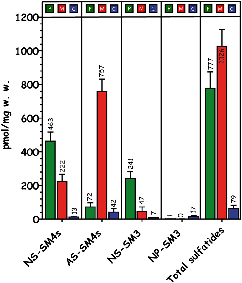
Fig. 3.
Quantitative distribution of renal sulfatides in the papillae (P), medulla (M), and cortex (C). Quantitative data were investigated by UPLC-ESI-MS2 (n = 3). The highest concentrations of sulfated GSLs were recorded in the medulla followed by the papillae (76% of the medulla) and more than 10-fold less sulfatide concentrations in the cortex (8% of the medulla). In the medulla, 74% of all sulfatides are AS-sulfatides, whereas in the papillae, 91% are NS-sulfatides. Finally the concentration of NP-sulfatides is 17-fold higher in the cortex than in any other region.

