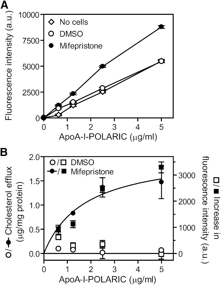
Fig. 2.
Detection of HDL formation by apoA-I-POLARIC in BHK/ABCA1 cells. BHK/ABCA1 cells were treated with or without 10 nM mifepristone for 20 h and incubated with the indicated concentrations of apoA-I-POLARIC for 6 h. A: Fluorescence intensity of the medium was determined. No cells (open diamonds), DMSO-treated cells (open circles), mifepristone-treated cells (closed circles). B: Cholesterol efflux from DMSO-treated cells (open circles) and mifepristone-treated cells (closed circles) was determined. Increase in fluorescence intensity of apoA-I-POLARIC was calculated by subtracting the value of no-cells from that of DMSO-treated cells (open squares) or that of mifepristone-treated cells (closed squares).

