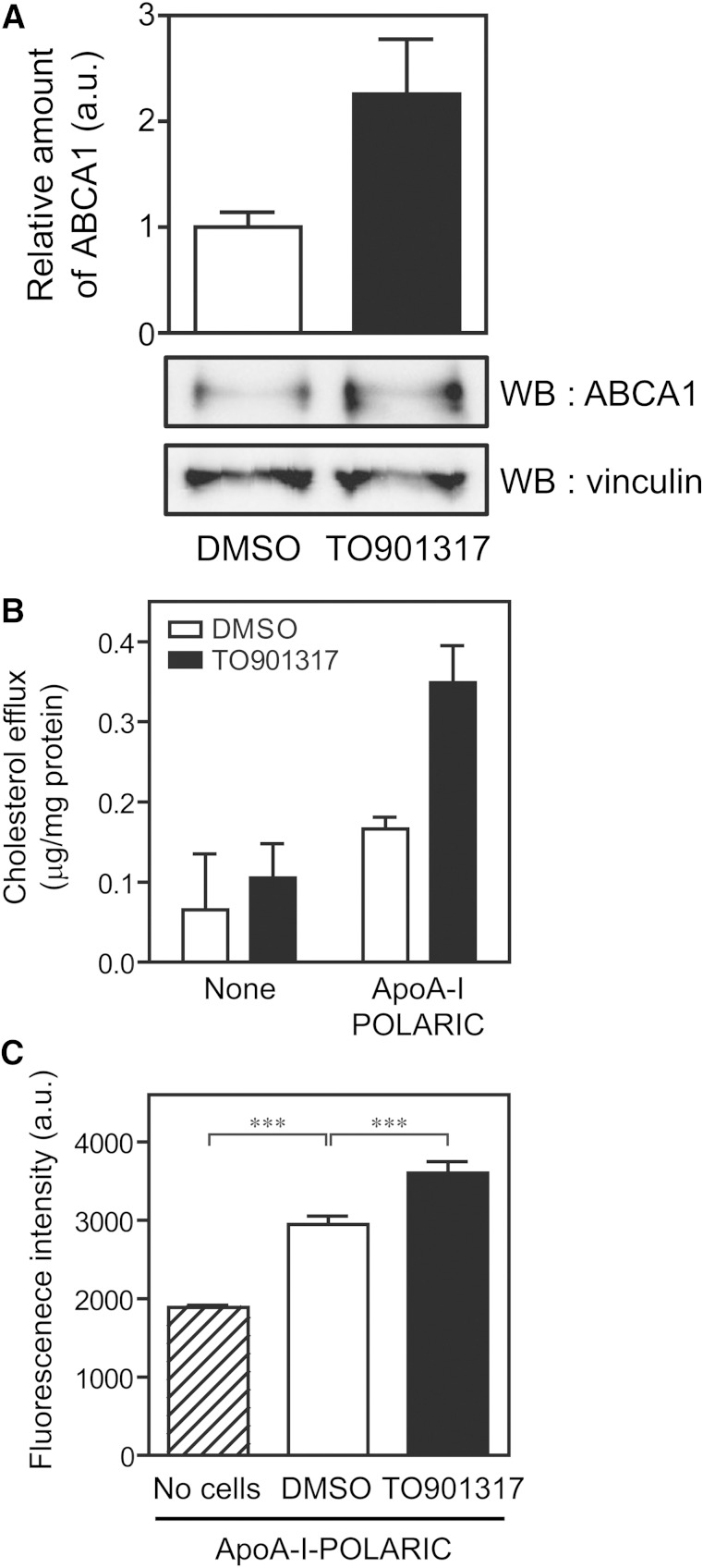
Fig. 4.
Detection of HDL formation by apoA-I-POLARIC in THP-1 macrophages. A: THP-1 macrophages were treated with 10 μM TO901317 for 24 h to induce the expression of ABCA1. Cell lysates (10 μg) were separated by 7% polyacrylamide gel electrophoresis, and ABCA1 and vinculin (loading control) were detected using the indicated antibodies. The amount of ABCA1 was normalized against vinculin. B, C: THP-1 macrophages were treated with (filled bars) or without (open bars) 10 μM TO901317 for 24 h and incubated with or without apoA-I-POLARIC (2.5 μg/ml) for 6 h. Cholesterol content in the medium (B) and fluorescence intensity of the medium (C) were determined (n = 3). ***P < 0.001; WB, Western blot.

