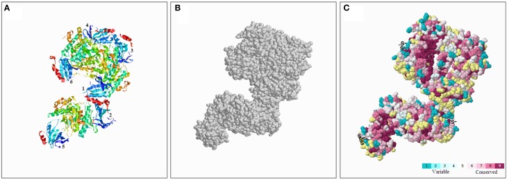
Figure 5.
Conserved evolution of SnRK2.6b—3D. (A) Crystal structure of the putative SnRK2 protein based on SnRK2.6b. The six subunits are marked with 1 and 6, respectively. (B) The amino acids were presented as gray balls on the spatial structure. (C) Residues are colored according to their levels of conservation. The score is 1–9 as blue to purple, which represents viable to conserve.

