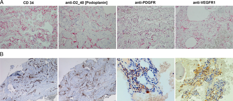
FIGURE 1.

Immunhistochemistry of pleural biopsy in case 1 (A), and of left ileosacral soft tissue in case 2 (B). Formalin-fixed, paraffin-embedded archival tissues were processed using standard IHC methods and stained with anti-VEGFR-1, anti-PDGFR, and anti-podoplanin (D2-40) antibodies (all from ZYTOMED Systems; GmbH) at 1:20 dilution. In both cases the CD34+ endothelium was also positive for D2-40 confirming its lymphatic origin. Further support for the therapy was the positive staining for PDGFR and VEGFR-1 tyrosine kinases, both targets of sunitinib. All images were taken at ×20 magnification with the exception of PDGFR and VEGFR in case B (ileosacral soft tissue), which was captured with ×40 lens. VEGFR indicates vascular endothelial growth factor receptor.
