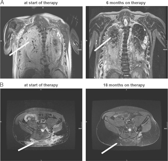
FIGURE 2.

Radiologic findings. For both patients T2-weighted magnetic resonance imaging at baseline (before initiation of therapy), at 6 and 18 months on therapy. In both, the thorax images for case 1 (A), and the pelvis images in case 2 (B) there is a marked reduction of the pathologic lymphatic tissue burden, as well as of the accompanying edema.
