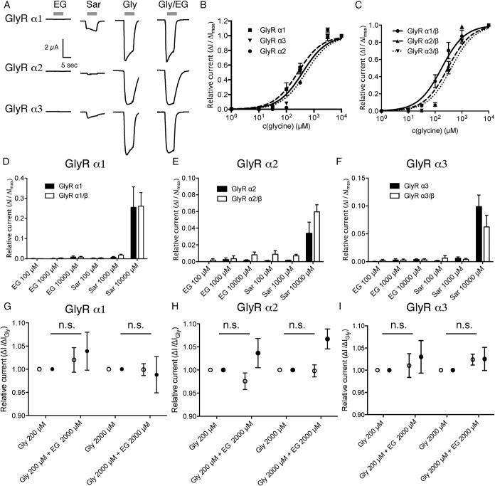
Figure 2.
N-ethylglycine (EG) does not induce significant glycine receptor (GlyR)–mediated currents in Xenopus laevis oocytes. Xenopus laevis oocytes were injected with complementary RNA encoding for mouse GlyR α1, GlyR α2, or GlyR α3 alone or coinjected with complementary RNA encoding for GlyR β. Substance-induced currents were recorded with the membrane potential held at −50 mV. The respective substances were applied for 15 seconds each followed by a washout of 30 seconds. (A), Representative substance-induced currents for 200 μM glycine (Gly), 1 mM sarcosine (Sar), and 1 mM EG were recorded from oocytes expressing the indicated GlyR subunit. (B and C), Concentration/response curve of currents elicited by glycine normalized to maximal glycine inducible currents as determined on oocytes expressing GlyR α1, GlyR α2, or GlyR α3 (depicted in B) or GlyR α1/β, GlyR α2/β, or GlyR α3/β (depicted in C), respectively. Data are expressed as mean ± SEM (n = 4-6). (D-F), Oocytes expressing GlyR α1 (D), GlyR α2 (E), or GlyR α3 (F) or the respective heteropentameric GlyR α/β receptors were superfused with EG- or sarcosine-containing solutions. Substance-induced currents were given in relation to the maximal glycine-induced current. Note that although small sarcosine-induced currents were observed in recordings from oocytes expressing any of the GlyR subunits, no or negligible EG-induced currents were observed. Data are expressed as mean ± SEM (n = 4-6). (G-I), Oocytes expressing the respective GlyR subunit (GlyR α1 in [G], GlyR α2 in [H], or GlyR α3 in [I], depicted with open circles) or the respective heteropentameric GlyR α/β receptors (depicted as filled circles) were superfused with glycine at the indicated concentration alone or in combination with 2 mM EG. Currents were determined as fraction of the current elicited by glycine alone. Data are expressed as mean ± SEM (n = 4-6).

