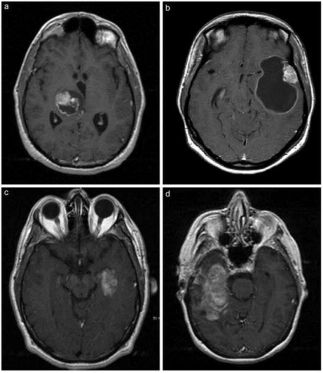
Figure 1.

A: Axial T1 post-contrast MRI shows a heterogeneously enhancing cystic mass in the right thalamus with mass effect but minimal surrounding vasogenic edema. Patient #6 illustrated.
B. Axial T1 post-contrast MRI shows an enhancing mural nodule in the left lateral temporal cortex associated with a large rim-enhancing cyst. Patient #9 illustrated.
C. Axial T1 post-contrast MRI shows an inhomogeneously enhancing lesion in the mesial left temporal lobe. Patient #4 illustrated.
D. Axial T1 post-contrast MRI shows a large heterogeneously enhancing lesion with complex cystic component within the right temporal lobe. Note the paucity of mass effect and the relative absence of vasogenic edema. Patient #2 illustrated.
