Figure 2.

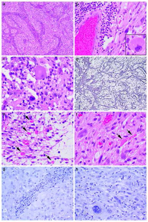
Direct comparison between PXA areas in Patient #1 (a, c, e, g) (a 27-year old female with relatively separate classic PXA and high grade glioma areas of tumor in whom no BRAF mutation was detected yet is a 10+ year survivor) versus Patient #3 (b, d, f, h) (a 68 year-old female also with relatively separate classic PXA and high grade glioma areas in whom BRAF mutation was detected yet survived less than 2 years). Note typical features of PXA including brisk non neoplastic lymphocytic collections (a, b), large pleomorphic cells (b inset, c), focally increased reticulin fibers (d), eosinophilic granular bodies (arrows, e, f) and negative immunostaining for IDH1 (g, h). Hematoxylin and eosin (a 100×, b 400×, b inset 600×, c 600×, e 600×, f 600×), reticulin stain (d 200×), and immunostaining for IDH1 with light hematoxylin counterstain (g, h, both 400×).
