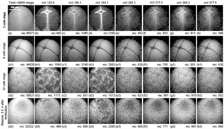
Fig. 7.
Total ion and selected ion images (positive mode ToF-SIMS) of the animal hemisphere of X. laevis embryos in different cleavage stages using dynamic ToF-SIMS imaging. The 40 keV C60+ beam primary ion dose density was 2 × 10¹³ ions/cm2 over a 1,200 × 1,200 μm2 area with 256 × 256 pixels. The biomolecular arrangements on the animal hemisphere of embryos at 2 cells, 4 cells, 32 cells, and blastula 5.5 h postinsemination are shown. During the cleavage, the embryo does not grow but divides into smaller cells (blastomeres) packed on the surface of the sphere. The maximum counts (mc) can be read on the ion images. The m/z 165.1, common fragment from glycerophospholipids; GPCho and SM share the fragments at m/z 125.0, 166.1, 184.1, and m/z 265.2 is unique to SM; m/z 313.3, MAG [C19H37O3]+ C16:0; m/z 369.4, cholesterol fragment; m/z 577.5, DAG [C37H69O4]+ C34:1 or the fragment from phospholipid. [Data previewed from (45)].

