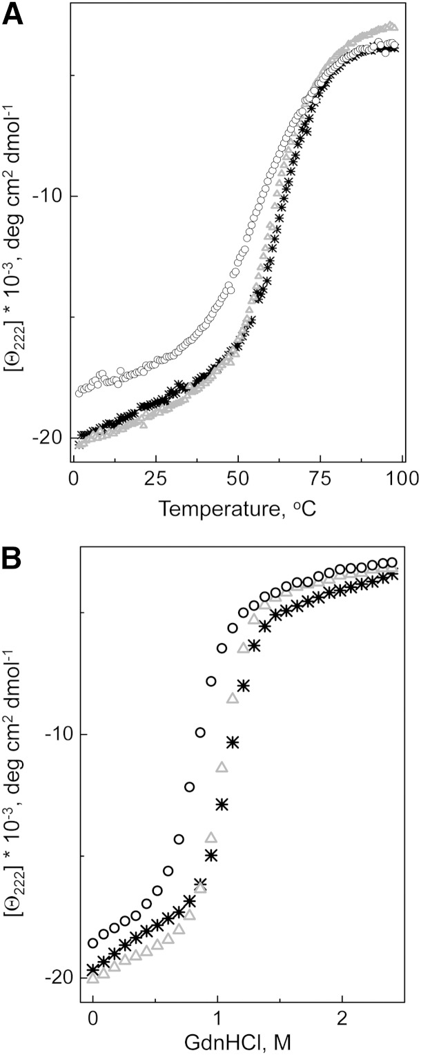
Fig. 5.
Unfolding of the lipid-free WT apoA-I and the apoA-I variants monitored by ellipticity at 222 nm. A: Thermal unfolding was induced by heating the protein from 2 to 98°C in the cuvette within the CD spectrometer holder. B: Chemical unfolding was induced by adding GdnHCl, in 0.1 M increments, to the protein inside the cuvette within the CD spectrometer holder. *, WT apoA-I; Δ, apoA-I[K107A]; ○, apoA-I[K107del].

